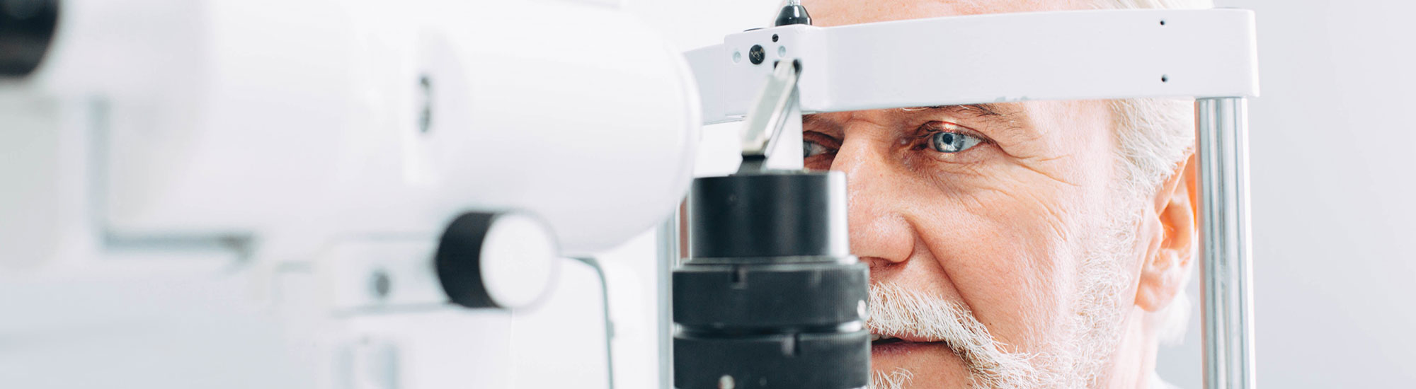
Adult & Senior Comprehensive Eye Exam Morden
Understanding Our Eye Health Assessment Process
Your eyes tell you a lot. They tell your optometrist how well you should see but also aspects of your overall health.
- Your eye exam will begin with a series of pre-tests to learn as much as possible about your eyes.
- Your optometrist will review the data before completing your comprehensive assessment with you.

A trusted relationship with your eye doctor is the best way to monitor changes over time. It’s our privilege to be your partner in eye health.
Detailed Pre-test
Our team will carry out a series of detailed tests to help us get as much information as possible about your eyes before you see your optometrist. Please click on the link for a detailed description of each.
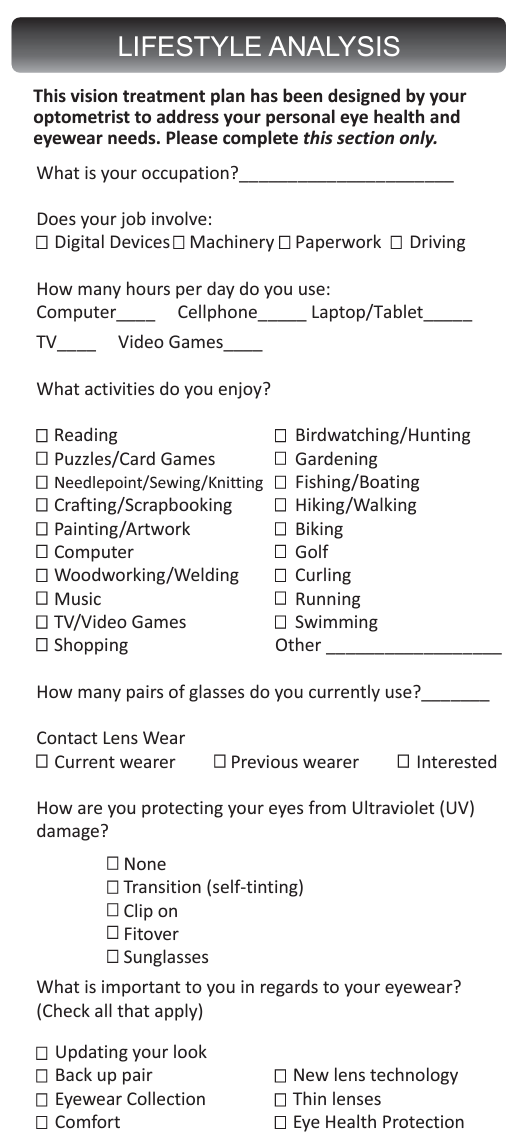
Through lifestyle analysis your optometrist and eyewear consultants can determine the perfect lens for your needs and prescription allowing you to experience optimal vision and function.
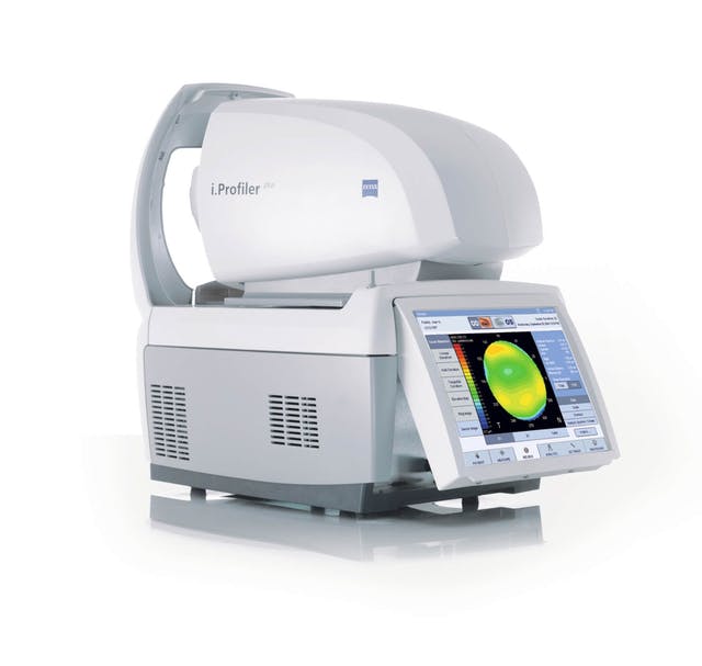
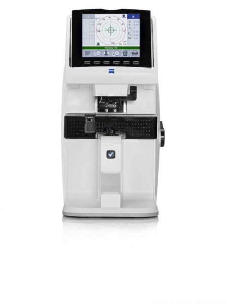
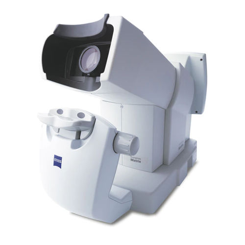
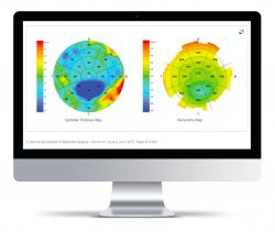
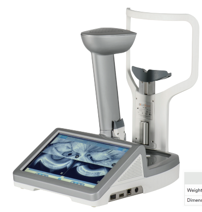
With this digital technology your prescription can be measured to 1/100th of a dioptre, offering crisper, brighter vision, especially at night. The iProfiler Plus takes into account your unique “eye print” (corneal map) which enables your optometrist to prescribe the most precise, balanced prescription for you. The corneal map or topography is a three-dimensional image of the surface of the cornea, the clear tissue at the front of the eye. Having this information is helpful in diagnosing corneal diseases such as Keratoconus earlier.
Measuring and verifying the prescription of your current eyewear is important to ensure that they are exactly as ordered and to monitor prescription changes. The Lensometer is also used to measure how well your current sunwear are blocking ultraviolet light.
Non-Contact Tonometer (NCT):
This instrument measures the internal pressure of your eyes known as the Intraocular Pressure (IOP). Pachymetry, a measurement of your corneal thickness, can be obtained at the same time on our instrument. This measurement is required to ensure that the pressure readings are within range for your given eye anatomy. IOP measurements are an important part of identifying inflammation and glaucoma risk. This test is particularly important because you have no internal sensation to tell you if your eye pressure is abnormal.
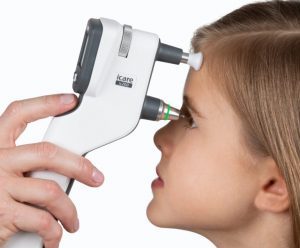
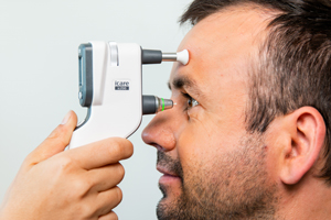
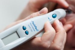
iCare Tonometer:
This “puff-less” instrument also measures your internal eye pressure (IOP). It measures your IOP without the puff of air, as with the non-contact tonometer. You can request this test over the “puff of air” test if you prefer.
The FDT measures your central and side vision using a variety of light patterns. It has been instrumental in detecting early eye disease and neurological (brain) disease.
Ishihara Colour Test
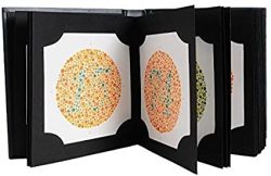
The Ishihara colour test, which consists of a series of pictures of colored spots, is the test most often used to diagnose red–green color deficiencies.
Farnsworth D15 Colour Test
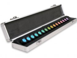
A more in-depth test that can be used is the Farnsworth D15. Each D15 set contains a reference disc and fifteen numbered discs, which make up an incomplete color circle. Following an attempt to sequentially arrange the discs by the patient, evaluation determines red-green or yellow-blue deficiencies.
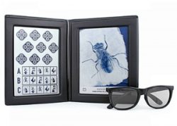
Stereo Fly
Stereoacuity is a measure of fine depth discrimination. This test has proven to be an effective and easy-to-use method of screening vision for all ages. They help to identify vision problems and conduct stereopsis, amblyopia, suppression, and strabismus testing, each of which can impede a child’s development and performance.
Pachymeter
Corneal pachymetry is the process of measuring the thickness of the cornea. A pachymeter is a medical device used to measure the thickness of the eye’s cornea. It is useful in screening for patients suspected of developing glaucoma among other uses.
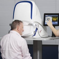
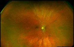
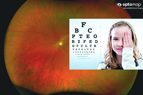
The Optomap non-invasively captures an instantaneous, ultra-widefield digital scan of the retina, revealing important information for the comprehensive evaluation of systemic and ocular health. Using the 200⁰ image of the back of your eye, the retina, your optometrist is easily able to detect any abnormalities. It also takes a scan using autofluorescence. Fundus autofluorescence or FAF imaging gives information over and above conventional imaging techniques such as fundus retinal photos as it allows visualization of retinal metabolic changes and helps to identify areas that may be at high risk for certain eye diseases. Optomap and FAF have been so integral to the diagnosis of eye pathology, Focal Point has made it a standard of care in our practice.
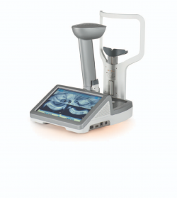
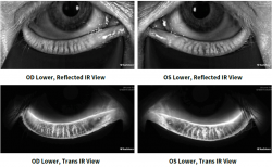
This non-invasive infrared camera images the meibomian glands. Meibomian glands sit within your eyelids and produce oils which form the outermost, or top, layer of your tears. Damage to, or loss of, these glands can lead to dry eye disease.
Insert information
Adult and Senior Eye Exam
After your pre-tests, your optometrist will use the detailed data to complete your comprehensive vision and eye health exam.
Your optometrist will evaluate how well you are seeing with and without glasses. When you have two eyes, teamwork is important. Your optometrist will examine how well your eyes work together and how they process visual information (colour, depth).
Using a phoropter your optometrist will determine the combination of lenses that will provide the best visual acuity
Using a biomicroscope and other hand help instruments your optometrist can view the external and internal structures of your eye.
Many eye and vision problems have no obvious signs or symptoms, so you might not know a problem exists. With a full view of your ocular health, your optometrist can detect problems earlier. Early diagnosis and treatment of eye problems can help prevent vision loss.
Additional specialized testing may be required if a health issue is suspected.
Child Eye Exams
The Canadian Association of Optometrists recommends that babies have their first eye examination between six and nine months of age. This is an important time to ensure eyes are healthy, eye and muscle movements and alignment are developing properly, and the eyes are focusing together. An accurate and complete eye exam can be performed without the need to read the eye chart. Your optometrist will test for nearsightedness (myopia), farsightedness (hyperopia), astigmatism, eye movement, and eye health.
There are a few ways to make this first visit more enjoyable:
- Work around fussy times.
- Schedule the appointment at a time when baby is generally relaxed and happy
By age 3, your optometrist should reassess your child’s visual system to confirm the absence of any eye disease, as well as monitor the continued growth and efficiency of visual skill development. This is also the examination where eye muscle problems such as crossed-eyes (strabismus) and lazy eye (amblyopia) are carefully assessed.
Tests for Children
Your optometrist and our team will carry out a series of detailed tests to help us get as much information as possible about your child’s eyes. If your child is able, these may include:
Your optometrist will evaluate how well your child’s eyes are seeing with and without glasses. When you have two eyes, teamwork is important. Your optometrist will examine how well your child’s eyes work together and how they process visual information.
Using a biomicroscope and other hand help instruments your optometrist can view the external and internal structures of your child’s eyes
Focal Point has made the Optomap retina scan free of charge for any child’s first scan. It can help spot peripheral retinal issues that can be missed as not all children can cooperate during the optometrists retinal exam. This initial scan provides a baseline retinal image to compare as your child grows. We encourage you to continue with future Optomap scans as we monitor your child’s eye health and vision.
The information from the autorefractor gives your optometrist a good idea of what your vision needs are. When they ask you to look at a chart through different lenses, they’re further refining their understanding of how well you see. The autorefractor gives them a good idea of what lens strength to offer you. This isn’t always accurate or possible with children which is why your optometrist performs retinoscopy. Retinoscopy is a manual method to determine one’s prescription. It involves shining a light into your eye and based on the pupil reflex the optometrist can determine what your prescription is. To further increase the accuracy it is common to dilate children before checking their prescription.
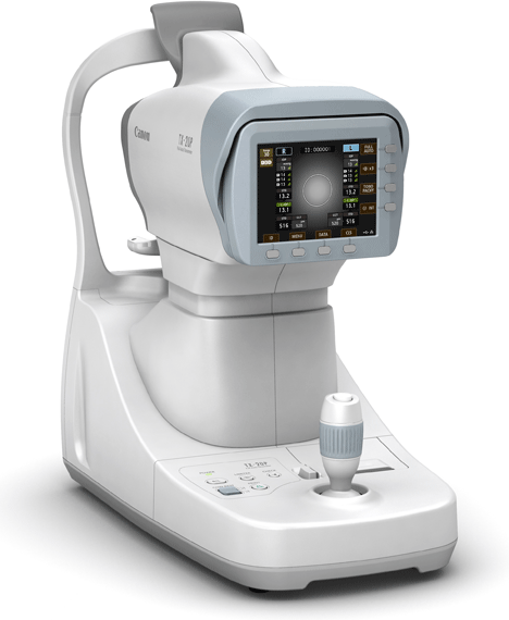
The instrument also measures the shape of the central cornea (keratometry).
This “puff-less” instrument also measures your internal eye pressure (IOP). It measures your IOP without the puff of air, as with the non-contact tonometer. You can request this test over the “puff of air” test if you prefer.
The FDT measures your central and side vision using a variety of light patterns. It has been instrumental in detecting early eye disease and neurological (brain) disease.
This non-invasive infrared camera images the meibomian glands. Meibomian glands sit within your eyelids and produce oils which form the outermost, or top, layer of your tears. Damage to, or loss of, these glands can lead to dry eye disease.
Stereoacuity is a measure of fine depth discrimination. This test has proven to be an effective and easy-to-use method of screening vision for all ages. They help to identify vision problems and conduct stereopsis, amblyopia, suppression, and strabismus testing, each of which can impede a child’s development and performance.
The Ishihara color test, which consists of a series of pictures of colored spots, is the test most often used to diagnose red–green color deficiencies.
A more in-depth test that can be used is the Farnsworth D15. Each D15 set contains a reference disc and fifteen numbered discs, which make up an incomplete color circle. Following an attempt to sequentially arrange the discs by the patient, evaluation determines red-green or yellow-blue deficiencies.